Fundamentals of Sound
MODULE 3: HEARING
Physiology / Function


The Ear as a Transducer - Introduction / Outer & Middle Ears
Transduction in the Inner Ear
           The Basilar Membrane - Auditory Interference & Masking - Nonlinearities
Optional Sections Detailing Auditory Function


THE EAR AS A TRANSDUCER - Introduction / Outer & Middle Ear


(source)

Transducers: Devices that convert

a) physical/chemical energy into electrical signals; e.g. sensors (mechanical, chemical, photoelectric etc.),
 
b) electrical signals into physical/chemical energy; e.g. actuators (robots, electric motors, etc.), or
 
c) both (a) and (b); e.g. bidirectional (voice coils: sensors in microphones, actuators in loudspeakers; antennae; etc.).

The ear is generally thought of as a sensor (e.g. a microphone) but it actually is a bidirectional transducer
 
Interesting Facts About the Ear:

  • it is surrounded by the hardest bone in the human body (temporal bone), necessary to protect the very fragile and important structures it contains, and insulate them from the various vibrations/sounds generated within the body;
     

  • it contains the smallest and lightest bones in the human body (ossicles, within the middle ear); and
     

  • it is the only organ that includes an amplifier (cochlea, in the inner ear), and the only sensory organ that is fully functional before birth (at ~25 weeks of gestation).

TRANSDUCTION PROCESS IN THE OUTER AND MIDDLE EAR

OUTER EAR

Overall Functions

_ concentrates and funnels sound wave energy from the air towards the middle ear;
 
_ protects the sensitive entrance to the middle/inner ear (eardrum);
 
_ selectively amplifies frequencies that are significant to the human voice.

Main Parts & Functions

  • Pinna or auricle (external part of the outer ear): a funnel-like appendix that receives sound waves generated by a source
    and propagated through the air.
     
    Functions
    :
     
    _ its various folds and crevices act as resonators:
     
           the larger ones amplify frequencies important to speech (~1-6kHz);
     
           the smaller ones, or more specifically, differences in the smaller ones between our left and right ear amplify high
           frequencies differently, providing us with spectral cues about where a sound is coming from
           (more during the "sound source localization" module);
     
    _ the entire pinna helps funnel and concentrate wave energy from the outside into the ear through the narrow ear canal
       (
    or auditory canal or auditory meatus)
     

  • Auditory canal: an ~2.5cm-long, almost cylindrical tube that receives sound waves entering through the pinna (it reaches its final length by age 7).
     
    Functions:
     
    _ it protects the eardrum, found at the far end of the narrow canal, from physical damage;
       hair and wax that line the canal further protect the eardrum from smaller particles and organisms
     
    _ it directs wave energy entering via the pinna towards the tympanic membrane or eardrum, setting it into motion;
       because of its shape/size (0.025m long cylinder, open on one end), it resonates at a range of frequencies (1-6kHz)
       that amplify vocal signals.

NOTE: Given the hard, temporal bone surrounding the entire hearing apparatus, sound energy can reach the inner ear not only through air conduction (i.e. by going through the ear canal) but also thought bone conduction, by applying sonic vibrations directly onto the skull. Bone conduction headphones work on this principle, applying vibrations directly onto the bone, behind the pinna.
 
(Optional: History of bone conduction technology in hearing and list of select available bone conduction headsets)

Simplified graph of the ear: in anatomical context (left) and magnified & sectioned (right)

    From "Introduction to Psychology," Walden University

Schematic: Longitudinal waves reaching the eardrum                                  (enlarge - animation)


MIDDLE EAR

Overall Functions

_ converts air pressure variations into mechanical vibrations, without in any way altering an incoming signal's frequency and time profiles (i.e. without altering its spectral and signal envelopes);

_ reduces the impedance mismatch between the outside (air in the outer ear) and the inside (liquid in the inner ear) of the ear, so that a sufficient portion of sound-wave energy from the outside can enter the transduction mechanism in the inner ear and be converted into electrical messages (rather than be reflected back outside).

Main Parts & Functions

  • Tympanic membrane or eardrum: a very thin, delicate, stretched, roughly circular membrane, ~10mm in diameter, that receives sound waves through the ear canal.
     
    Functions:
     
    _ converts acoustic energy (sound waves) into mechanical energy (membrane vibrations)
     
    _ it is attached to the first (malleus or hammer) of three interconnected bones (ossicles) in order to transfer its mechanical
       vibrations to another membrane at the entrance of the inner ear, the oval window, which is ~20 times smaller in area; this
       area decrease increases the pressure by 20 times and helps reduce the impedance mismatch between the outer & inner ears.
     
       [Optional] This mismatch is further reduced through the "buckling" or "stir-like" motion of the eardrum.
        This motion reduces the eardrum's vibration velocity, particularly for high frequencies, and increases
        in turn the force with which the eardrum pushes on the ossicles (conservation of momentum).]


  • the ossicles are three delicate, interconnected bones [malleus (hammer), incus (anvil), stapes (stirrup); lightest bones in the body],  arranged into a lever-like formation; the first (hammer) is attached to the eardrum and the last (stirrup) is attached to the oval window, at the entrance to the inner ear.
     
    Functions:
     
    _ their lever arrangement and area difference between eardrum and oval window reduce the impedance mismatch
       between the outer and inner ears:
        
            the lever arrangement (the hammer is ~1.3 times as long as the anvil) reduces the displacement and velocity of the oval window,
            relative to that of the eardrum, and consequently (due to conservation of momentum) increases the force with which the stirrup
            pushes onto the oval window and into the inner ear by a factor of ~3-4;
     
            as noted previously, the ossicles transfer the eardrum's mechanical vibrations to another membrane at the entrance to
            the inner ear, the oval window, which is ~20 times smaller in area; this area decrease increases the pressure by ~20 times
            and helps further reduce the impedance mismatch between the outer & inner ears.
       
            so: ~3-4 times increase in force due to the level mechanism x ~20 times increase in force due to transfer to a smaller area
                  ~ 70 times increase of force thanks to the middle ear, facilitating vibration transfer from air (outer ear) to liquid (inner ear).
     
    _ they help dampen high level signals and protect the inner ear via two tiny muscles attached to them.
     
    [Optional: Similarly to the outer ear, but to a lesser degree, the middle ear (e.g. length and motion of the ossicles) also gives some preference (i.e. presents less resistance) to frequencies relevant to vocal signals.]

The air cavity of the middle ear can connect to the outside via the Eustachian tube, a passage that, when open, functions as an air pressure equalizer between the outer and middle ears (i.e. equalizes the pressure at the two sides of the eardrum). If the air pressure is not equalized, the eardrum's sensitivity drops and its response becomes non-flat (i.e. different sensitivity at different frequencies).


(a)                                                     (b)                                                     (c)                                                        (d)

(a) Eardrum motion & (b) Ossicle motion animations
(Wada Laboratory of Genetic Engineering; Tohoku University, Japan - site temporarily down).
Note the different modes of vibration on the membrane and the ossicles at high versus low excitation frequencies.
 
(c) [Optional] Animated eardrum motion data, measured on the ear of an American bullfrog (Physiological Science; UCLA).
(d) Detailed outline of outer/external and middle ear functions (NeurOreille, France).


TRANSDUCTION PROCESS IN THE INNER EAR


INNER EAR

Overall Functions

_ performs a spectral analysis at short, consecutive time windows on the incoming signal, breaking it down into its sinusoidal
   frequency components;
 
_ compresses the incoming dynamic range to enhance low-level signals and reduce high-level signals;
 
_ converts the resulting spectral information into electrical signals and conveys them to the brain.
 

Main (hearing-related) Parts & Functions

The hearing portion of the inner ear consists of the Cochlea, a small, snail-like structure (~9mm in diameter and ~5mm in height) that is split into three liquid-filled compartments (see the pictures, below):

_ The Top Compartment (scala vestibuli).
   ... is filled with a fluid (perilymph - neutral) that receives the middle-ear vibrations through the oval window,
   at the cochlear base. These vibrations ascend as pressure waves towards the apex (top end) of the cochlea
   and are passed on to the length of the middle compartment via conductive resonance;
 
_ The Middle Compartment (scala media).
   ... is filled with a different kind of fluid (endolymph - positively charged) that receives the scala vestibuli vibrations via a thin
  
membrane separating the two scalas (Reissner's membrane) and passes them on to the ear's spectral analyzer and
   transducer: the Organ of Corti (OofC).
 
          At the bottom of the OofC is a loosely-coupled collection of filaments, called the basilar membrane (BM), which performs
          spectral analysis on the incoming signals. The top portion of the OofC performs the transduction (see further below for
          details on the OofC and the BM)
 
_ The Bottom Compartment (scala tympani)
   ... communicates with the top compartment via an opening at the cochlear apex (top end), called helicotrema, and is filled with
   the same neutral fluid (perlilymph). It receives excess vibrations from the top compartment or scala vestibuli (i.e. vibrations
   not absorbed by the middle compartment or scala media), releasing them out of the cochlea through a small circular
   membrane called round window.


 ←---------------| 

 Top & Middle: two simplified schematics of a stretched-out cochlea;
The middle one marks the three cochlear compartments
Bottom: coarse simulation of the middle/inner ear action

Above Left: Microphotographs of two intact cochleae and of a dissected one.
The top one marks the oval and round windows.
The bottom one marks the Organ of Corti, which transduces mechanical energy into electrical neurological signals,
bringing auditory stimuli to the brain (see below).


Schematic cross-sections of the cochleae cochlea

Illustrations of the three key compartments or scalas (scala: stairs) and the main transduction element: the Organ of Corti.


(source)


Schematic close-up of the Organ of Corti

The Organ of Corti (OofC) is bounded
_ at the bottom by a collection of loosely-coupled elongated filaments, called the Basilar Membrane (BM), responsible for spectral analysis, and
_ at the top by a soft gelatinous tissue, called the Tectorial Membrane (TM), responsible for the mechanical initiation of the inner ear's mechanical-to-electrical transduction.
[Optional: Video of the transduction process in the Organ of Corti]

Transduction Process in the Organ of Corti - Summary

The BM (green line on the OofC cross-section animation, below) performs a spectral analysis by resonating at different places for different frequencies: high frequencies near the base - low frequencies near the apex.

As the BM moves, it pushes cells that lay above it (red pillars in the animation, called hair cells) up against the TM (gray structure).
This causes the tiny hair-cell tips (or hair bundles or stereocilia) to shear against the TM.
Inner Hair Cells (one row - tips not embedded on the TM) generate electrical impulses that encode the incoming waves' characteristics (the frequency and amplitude of its spectral components).
Outer Hair Cells (three rows - tips embedded on the TM) modify the movement of the TM to enhance low-level signals and reduce high-level signals.
 
The impulses travel along the auditory nerve pathways to the brain, entering a complex electrochemical network where the sensation of sound is registered. [Optional: short video on how this happens.]

As already noted, excess fluid vibrations, not absorbed by the BM, reach the scala tympani portion of the cochlea via the helicotrema (passage from scala vestibuli into scala tympani at the apex (far end) of the cochlea), and exit the cochlea through the the round window

Inner-Ear Innervation

Healthy (top) & damaged (below) hair cells
(from Curtis, 1979).

Exposure to high intensity sounds can result in

  • temporary hair cell damage, referred to as Temporary Threshold Shift or TTS (: temporary reduction in hair cell sensitivity) or

  • permanent hair cell damage.

We will return to Hearing Loss and Conservation during the "Loudness" module.

• ~30,000 auditory nerve fibers (neurons) are linked to auditory hair cells
 
• ~ 90-95% of the nerve fibers are linked to the inner hair cells, whose main function is to send messages to the brain about the frequency and amplitude of the incoming sound's spectral components;

1 inner hair cell may be connected with up-to 20 nerve fibers, most of which are afferent (sending messages from the ear to the brain).

• ~ 5-10% of the nerve fibers are linked to the outer hair cells, whose main function is to compress the level of the incoming signals by increasing response to low level signals and reducing response to high level signals.

Up to 10 outer hair cells are may be connected to 1 efferent nerve fiber (sending messages from the brain to the ear to support the OHC's amplification action).

Nerve Fiber's Spontaneous Activity: Firing activity of a single nerve fiber(neuron) in the absence of a stimulus. 
It determines the neuron's sensitivity and readiness to respond.
_ Neurons with higher spontaneous activity respond to lower level signals
_ Neurons with lower spontaneous activity respond to higher level signals.
 
Nerve Fiber's threshold: the minimum stimulus level that will increase the neuron’s firing rate above the spontaneous activity rate.  

Spontaneous activity discharge rate (number of electrical discharges/unit time): it ranges from 0 to 100 spikes/second. Maximum activity discharge rate: the theoretical maximum discharge rate of a neuron is ~ 1000 spikes/sec, although most auditory nerve fibers measured have discharge rates that max out at ~ 500 spikes/sec.


Videos outlining the auditory transduction process

Additional Videos @ https://www.interactive-biology.com/physiologyvideos/ (videos 036-040)  

 
Comprehensive, succinct, multimedia-rich site on the
Hearing Mechanism: http://www.cochlea.eu/en/ear


THE BASILAR MEMBRANE
 
Auditory Interference & Masking
Other Nonlinearities


Basilar membrane (BM): A collection of interconnected, weakly-coupled, flexible fibers located at the basis of the Organ of Corti, in the inner ear (cochlea). 
It is a tuned resonator that analyzes complex waves into sinusoidal components.

The BM is organized tonotopically: it vibrates at different places in response to incoming waves of different frequencies, and with different amplitudes in response to incoming waves of different intensities.

The resonance range of the human BM and, therefore, the frequency range of hearing (i.e. absolute thresholds for frequency) extends from ~20Hz to ~20.000Hz (20KHz).
 
On average:
_ Frequencies below 20Hz sound as individual pulses with no definite pitch.
_ Frequencies above 20KHz are inaudible.

250Hz:
Boystown Research Hospital

1kHz:
Boystown Research Hospital

4kHz:
Boystown Research Hospital 


For high frequencies, the basilar membrane vibrates close to the entrance/base of the cochlea, just next to the oval window, where the membrane is thinner, narrower, & tenser/stiffer.

For low frequencies, the basilar membrane vibrates towards the apex (top end) of the cochlea, where the membrane is thicker, wider, & looser.

The BM moves more efficiently at places along its length corresponding to the frequency range associated with speech signals (1-6kHz).
 
The tips of tiny hair cells (nerve endings) on the organ of Corti are pushed against the Tectorial Membrane (TM) by the motion of the basilar membrane, translating this motion into electric impulses.

CRITICAL BAND vs CRITICAL BANDWIDTH

Critical band: Term introduced by Fletcher in the 1940s to refer to the frequency bandwidth of the, then loosely defined, auditory filter. Since von Békésy’s studies (1930s-1960s), the term refers literally to the specific portion of the basilar membrane that goes into vibration in resonance with an incoming sine wave. Its length is determined by the elastic properties of the membrane and, at middle frequencies, has an average value of ~1mm.

Critical bandwidth: the frequency difference (~ 1/3 of an octave) corresponding to the physical length of the critical band .
It can be defined as

  • the minimum frequency difference between simultaneous sine waves with comparable levels required for them to sound free of beating / roughness sensations (free of interference)
    or

  • the minimum frequency difference between two simultaneous sine waves with largely unequal levels required for them to sound as two distinct tones vs. the weak wave being masked (perceptually "covered") by the strong wave.

 
AUDITORY INTERFERENCE

If the frequency difference between two simultaneous sine waves with comparable levels is within the critical bandwidth, the ear will not be able to resolve the two frequencies and the waves will interact in specific and musically important ways:

_ If the frequency difference is  ~< 10-20 Hz, the wave interaction will be perceived as a slow loudness fluctuation called beating
 
_ If the frequency difference is  ~> 20 Hz  but smaller than the critical bandwidth, the interaction of the two simultaneous waves will be perceived as a change in the character of the combined sound referred to as roughness

 
As already mentioned, critical bandwidth may therefore be defined as the minimum frequency separation in Hz. between two simultaneous sine waves necessary for beats/roughness to disappear and for the resulting tones to sound clearly apart.
 
Both, the beating and roughness sensations are perceptual attributes of amplitude fluctuation resulting from sound wave interference (discussed previously).

Psycho-physiologically, the beating and roughness sensations are linked to
 
a) the inability of the auditory frequency-analysis mechanism to resolve inputs whose frequency difference is smaller than the critical bandwidth
and
b) the resulting instability or periodic “tickling” (Campbell and Greated 1987: 61) of the mechanical system (basilar membrane) that resonates in response to such inputs.
 
[We will return to beating and roughness, when discussing musical timbre, consonance, and dissonance.]

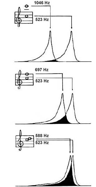
As the interval between two tones of comparable levels decreases, their respective disturbances on the basilar membrane (critical bands) increasingly overlap, resulting in the sensations of roughness and beating
(in Campbell & Greated, 1987).

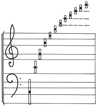
Harmonic components & Critical Bandwidth

The first 12 components of C3, shown as black circles on a stretched music-notation 'stave'.
The vertical bars indicate the approximate critical bandwidth around each component
Campbell and Greated, 1987).

Beating/Roughness Resources to review:

1) Read this description of the relationship among beating, roughness, spectral distribution, and critical bands.

2) Listen to a comparison between the roughness and beating sensations. As the lower-pitched tone in the interval  (i.e. note-pair) rises and the frequency difference between the tones gradually narrows from D5-Eb5 to Eb5-Eb5, the roughness sensation gradually gives way to the beating sensation.

3) Watch this clip, presenting two simultaneous sine tones of equal amplitudes. One is fixed at 1000Hz; the other can sweep between 600Hz and 1300Hz. When the sweep tone is within ~1-20Hz from 1000Hz, we have beating. For larger frequency differences between the sweep and the fixed tones we get various degrees of roughness.
Roughness disappears when the sweep frequency drops below ~800Hz or so, at which point when we hear two tones.
However, roughness persists for sweep tone frequencies past 1200Hz. This is because the critical bandwidth is larger in Hz for center frequencies of 1100Hz (two equally strong tones at 1000Hz and 1200Hz) than for center frequencies of 900Hz (two equally strong tones at 800Hz and 1000Hz). 


AUDITORY MASKING

Because the critical bandwidth (i.e. the frequency response range on the BM for a single frequency input) is rather broad (~1/3 octave at middle frequencies), tones separated in frequency by less than the critical bandwidth
a) will either interact to produce the sensations of beating and roughness (if they have comparable levels - see above), or
b) will result in masking (if they have different levels - see below).


Simultaneous Masking

Term describing the ability of one tone or band of noise (masker) to cover or raise the audibility threshold of a second tone (signal).

When two tones (or a band of noise and a tone) close in frequency are presented simultaneously, one (masker) may mask or “cover” the other (signal) depending on their level difference: the more intense tone may mask the less intense tone.
The smaller the frequency difference and the larger the level difference between two simultaneous tones, the more likely it is for masking to occur.

As the response of the BM around the characteristic (resonant) frequency is asymmetrical (larger above than below characteristic frequency), the resulting masking curves are also asymmetrical, with the lower frequency tone (or noise band) in a pair being more likely to mask (i.e. being a more efficient masker than) the higher frequency tone.
Low frequency tones are more likely to mask high frequency tones.
Listen to this example of masking asymmetry

Simultaneous masking may be due to

a) “Swamping” at the BM and/or nerve fibers
The masker produces a significant amount of activity in the auditory filters, “swamping” the signal information and making it undetectable

b) “Suppression”
The neural response to the signal is suppressed by existing neural activity at a different area on the basilar membrane


As the level of a signal increases, the BM response  increases in magnitude and width (inverted v-shaped lines, above). This means that stronger signals are able to cover increasingly stronger simultaneous signals and increasingly further removed in frequency.
 
Response width increases more above than below the stimulating frequency. This means that any signal is more likely to cover simultaneous, lower-level signals that are higher vs. lower in frequency.

Forward and Backward Masking (Temporal Masking)

During Forward Masking, a signal masks a tone that comes 0-200ms after it. Forward masking effects are slight and do not produce the broad masking effects of simultaneous masking.

Forward masking increases with masker duration (from 0 to 50ms) and with masker level. It occurs most effectively for masker-signal delays ~20-30ms and does not occur for delays >200ms.

Forward masking depends on the signals used and may be due to masker activity persisting at some level in the auditory system, impacting signal perception.

During Backward Masking, a signal masks a tone that came 0-50ms before it.
Backward masking effects are even slighter than those of forward masking and are more prominent in untrained listeners.

Backwards masking depends on the signals used and may be due to higher level cognitive processing.

Listen to this example of backward and forward masking


Review the Masking Slides presented in class.

Listen to two audio examples of noise bursts masking gaps in a steady or frequency modulated tone .
Listen to the steady and frequency modulated tones without the noise bursts; the gaps are now clearly audible (after Dannenbring,1974).

 
NONLINEARITIES OF THE EAR
Main "Side-effects":
Distortion - Suppression - Otoacoustic Emissions

Distortion
The BM introduces a variety of distortion products, related mainly to two types of distortion:
Harmonic Distortion
and Intermodulation Distortion

Harmonic distortion of a sinusoid stimulus will introduce frequencies that are integer multiples of (or harmonically related to) the stimulus frequency. Since frequency components that are harmonically related tend to fuse together into a single tone percept, harmonic distortion does not usually produce strong, undesirable perceptual effects.
 
Intermodulation distortion results from the interaction between two or more sinusoidal stimuli on the BM.  It introduces additional frequency components in the original tone's spectrum, which may be perceived as "combination" tones (i.e. tones not originally present, arising from the combination of the original tones). Intermodulation distortion products are often inharmonically related to the original tones and, consequently, rather noticeable.
[Optional: For a two-component stimulus with frequencies f1 and f2 (assuming f1 < f2), the most common intermodulation distortion products are: f2 - f1 (difference tone), f1 + f2 (summation tone), and 2f1-f2  &  2f2-f1 (2 of the 4 cubic distortion products)]

Suppression
Term describing the observation that the ear's response to one tone may decrease (i.e. may be suppressed) due to the presence of a second tone. Such suppression may occur at the BM or at neural levels.
 

Otoacoustic emissions

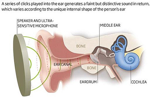
The ear acts not only as a microphone, receiving sound, but also as a speaker, emitting a series of tones referred to as otoacoustic emissions (OAEs). 

OAEs are classified depending on the context of emission (i.e. spontaneous vs. evoked and, if evoked, by what). 
The important thing to remember is that healthy/alive ears produce more of these emissions than unhealthy/dead ears, indicating that they may have something to do with the spontaneous activity of the nerve fibers attached to the inner/outer hair cells and the amplification effect of the outer hair cells.

Watch this video describing an application of OAEs to hearing assessment and headset tuning. Here is another relevant product.


[OPTIONAL SECTIONS]

What Types of Hair Cells and Nerve Fibers Populate the Inner Ear?

 
  • Inner hair cells. Their disturbance sends auditory messages to the brain down a complex auditory pathway, in the form of electrical impulses; afferent fibers carry information from the ear into the brain and are connected mostly to inner hair cells.
     
    Inner Hair Cell Action:

_ Basilar membrane motion pushes up against the organ Corti, resulting in shearing forces that bend the inner hair cell (IHC) stereocilia against the tectorial membrane.
 
_ IHC stereocilia tip links (protein filaments) stretch during bending, opening up ion channels in neighboring stereocilia
 
_ Positively charged Potassium (K) ions, flowing inside the endolymph that fills the scala media, enter the cells, attracted by the negatively charged (at rest) cells
 
_ The ensuing depolarization of the cells results in the release of neurotransmitters and the creation of neural activity corresponding to a series of electrical spikes.
 
_ The resulting signal to the brain is a rectified version of the stimulus signal because the cells fire only when the stereocilia bend towards the scala media, at the positive portions of the signal

Motion of the Organ of Corti and inner hair-cell action (Wada Laboratory, Japan - site temporarily down)

  • Outer hair cells. Their disturbance sends information to the base of the cell, influencing the cells' length in a feedback mechanism that helps the ear adjust its sensitivity based on the level of the incoming signal and on messages from the brain; efferent fibers carry information out of the brain into the ear and are connected mostly to outer hair cells. 
     
    Outer Hair Cell Action:

_ As is the case with IHCs, basilar membrane motion pushes up against the organ Corti, resulting in shearing forces that bend the outer hair cell (OHC) stereocilia against the tectorial membrane.
OHC stereocilia tip links (protein filaments) stretch during bending, opening up ion channels in neighboring stereocilia
 Positively charged Potassium (K) ions, flowing inside the endolymph that fills the scala media, enter the cells, attracted by the negatively charged (at rest) cells
 
_ Depolarization (occurring when the ion channels are open) and hyperpolarization (occurring when the ion channels are closed) of outer hair cells (OHCs) releases neurotransmitters that change the shape of the prestin protein molecules inside the OHCs, and, consequently, the OHC length, assisting in
a) amplification/attenuation, b) auditory filter sharpening, and c) compression of the ear’s dynamic response.
 
_ At low signal intensities, OHCs change periodically 900 out of phase with the TM movement, pulling on the BM, increasing IHC stereocilia bending, and amplifying the signal.
 
_ At high signal intensities, OHC change periodically in phase with the TM movement, pushing against the BM, decreasing IHC stereocilia bending, and attenuating the signal.
 
_ Whether there will be amplification or attenuation depends not on the length of the OHCs but on the phase relationship between OHC length changes and TM & BM vibrations.
 
_ In the short term, OHCs change in response to the bending of their own stereocilia. In long-term stimulation, OHCs may change in response to messages from the brain, carried to OHCs via efferent nerve fibers.


    (a)                                                        (b)                                             (c)                                                      (d)

(a) Basilar membrane vibration animations and (b) in-vivo (alive) vs. in-vitro (dead) vibration measurements
(Wada Laboratory of Genetic Engineering, Tohoku University, Japan - site temporarily down) 
 
(c)-(d) Detailed outlines of cochlear and Organ of Corti function (NeurOreille, France)


(a)                                                    (b)                                                    (c)

Outer hair-cell (a) motility, (b) explanation, and (c) amplification effect (Wada Laboratory, Japan - site temporarily down)
 

BASILAR MEMBRANE ENCODING OF FREQUENCY & AMPLITUDE

Temporal Coding:
Phase Locking and Rectification

Neural response (neural firing) follows (or appears to be locked to) the positive peaks in the stimulus, firing only when the stereocilia are sheared in one direction.

This results in the neural signals of sinusoidal inputs

i) having a pulse-like shape with repetition rate equal to the input's period (i.e. they represent frequency in the time domain) and
 
ii) being rectified (i.e. constricted into to the positive portion of the two-dimensional signal graph).

The process of phase locking is closely related to hearing's "temporal coding theory" of encoding frequency.

The inner hair cells release neurotransmitters only when the basilar membrane moves in one direction, towards the scala media. They therefore only respond to the positive portions of incoming signals, with their stereocilia opening their ion channels when bending against the TM, mostly at a signal’s positive peak.

Temporal coding assumes that, thanks to phase locking, the auditory system encodes periodic information through the firing rate of neurons. This rate corresponds to the incoming signal’s period (or some multiple of it) and, therefore frequency.
Since neurons are not fast enough to encode high frequencies, more than one neuron is involved in the process. Each neuron fires at some of the peak portions of an incoming signal and, after adding the outputs of all neurons, the signal is represented to the brain in a manner similar to that shown at the bottom graph (left).

Place Coding

The above figure illustrates an alternative to the "temporal coding theory" of encoding frequency, referred to as "place coding theory."

As we've discussed, the basilar membrane (BM) responds at different places for different frequencies, due to its mass-stiffness gradient.
Place coding assumes that the basilar membrane is a collection of overlapping bandpass filters.

In reality, the basilar membrane is a continuous array corresponding to much more numerous filters than those shown in the graph, above (24 in the Bark scale). High frequencies cause a response closer to the BM's base, while low frequencies cause a response closer to the apex. Frequency is then encoded by the IHCs that correspond to the resonating portion of the BM and share the same characteristic frequency with that portion.


The image to the left is a schematic diagram illustrating the signals sent to the brain when the basilar membrane is vibrating in response to a complex wave with many sine components.

Each low-frequency component sends individual signals since, as indicated above, the frequency separation between the low-frequency components is larger than the critical bandwidth.
The upper components/harmonics are 'unresolved' because, as the component number increases, more components fall within the same critical band (Campbell and Greated, 1987).


Overview Resources on Ear Anatomy and Function

Dangerous Decibels from the National Institute of Health website.
Although the materials are designed for younger audiences (high school students), they present a good basic overview of our topic.
Well-designed and concise overview of the anatomy and function of the ear, Part of NeurOreille's Journey Into the World of Hearing
The Auditory System: Structure and Function, part of the Neuroscience e-Textbook site at the Department of Neurobiology and Anatomy of the McGovern Medical School, University of Texas.
This overview goes into more detail, especially in terms of the inner ear.

Acoustics & Auditory Neuroscience Page - City University of Hong Kong (Table of Contents)

Interactive Sensation Laboratory Exercises (ISLE) - Hanover College, OH (Chapters 10-13)

Details on the nonlinearities of the inner ear - hearing module of a Psychoacoustics course

Everything about hearing, including the optional sections, in an 11-minute video!


Loyola Marymount University - School of Film & Television